No products in the basket.

The Role of Portable Bladder Scanners VS Portable Ultrasound Scaners Introduction to…
Read moreLoading
General medical US imaging machine supplier since 1980's
General medical US imaging machine supplier since 1980's
The Role of Portable Bladder Scanners VS Portable Ultrasound Scaners Introduction to…
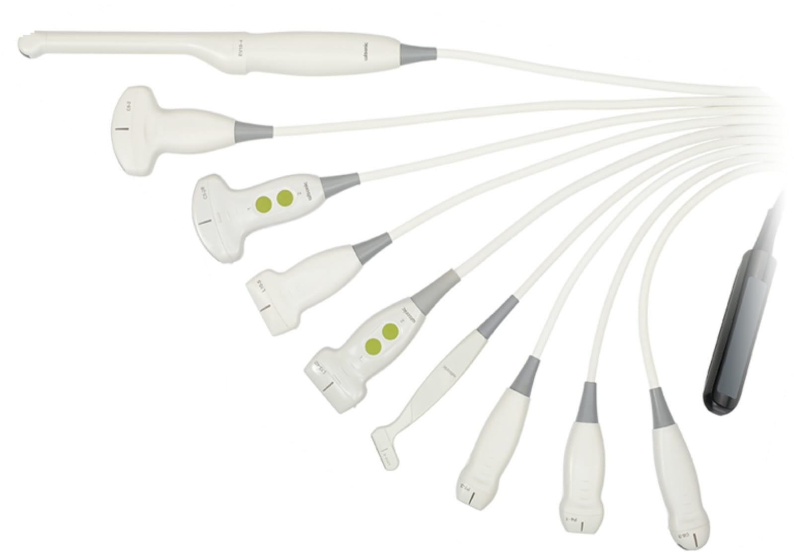
Read moreIntroduction to Ultrasound Transducers Ultrasound transducers play a pivotal role in the…
Read moreIntroduction to Ultrasound Governance In the realm of modern medicine, ultrasound has…
Read moreOverview of Ultrasound Imaging and Its Significance in Healthcare Ultrasound imaging, commonly…
Read moreIntroduction to Hand Held Ultrasound Scanners Hand held ultrasound scanners are reshaping…
Read more